

Source: https://enfermeriabuenosaires.com/caracteristicas
Characteristics and Functions
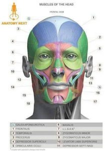
The muscles of the face are so numerous and varied that it can be very difficult to imagine how such a large number of muscle groups can be distributed in such a small space, and moreover, these muscles have such specific and differentiated functions.
The complexity of facial expression, in addition to the functions of phonation and swallowing, requires not only many muscle groups but also extraordinary coordination between the different muscle groups to achieve such a wide variety of movements, ranging from a simple wink to whistling.
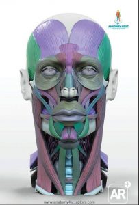
The muscles of the face can be divided into two main groups:
- Small size and with a function limited to facial expression,
- Large size with a fundamental role in important functions such as speech, eating, and even breathing.
General Characteristics
For the most part, they are flat, thin muscles with a rather discreet size/strength ratio; that is, they are not muscles with many muscle fibers or that generate great power.
Almost all of them have two insertions at different points, one in the facial bones and the other in the skin of the face or the aponeurosis of a neighboring muscle; sometimes both can be the case.
The larger muscles are an exception to this rule. These are the masseters, which have both their insertions on bone surfaces; they are capable of moving a joint and, per square centimeter of surface, are among the most powerful muscles in the body.
Classification and Functions
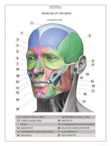
The facial muscles can be classified according to their function and according to the anatomical area with which they are related.
According to their function, the facial muscles can be divided into:
- Muscles of expression
- Muscles of mastication
The muscles of expression almost invariably insert into bone and skin, while those of mastication always insert into bony surfaces.
According to their anatomical location, the facial muscles can be divided into muscles of the:
- Auricles.
- Eyelids and eyebrows.
- Nose.
- Mouth and lips.
Muscles of the auricles
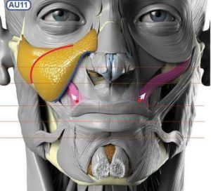
Due to the location of the auricles, their structure, and the particular evolution of humans—who no longer rely so much on hearing to survive—the muscles of the auricles are considered muscular vestiges.
Although they are present, their function is null. In fact, cases of individuals with the ability to move their auricles are exceptional.
In cases where it is possible to move them, it is due to the action of the anterior, posterior, and superior auricular muscles, which are present in all people but in very few cases have enough strength to have a visible effect.
Muscles of the eyelids and eyebrows
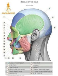
Their main function is to generate eyebrow movement, furrow the brow, and especially allow eye opening; this group includes the following muscles:
- Occipitofrontalis
- Procerus
- Corrugator supercilii.
- Orbicularis oculi.
The latter is the most important of all, as it allows eye closure; it is a large, flat, and circular muscle that surrounds the outer part of the orbits.
It is divided into several portions that allow the eyes to close gently or "squeeze" the eyes shut.
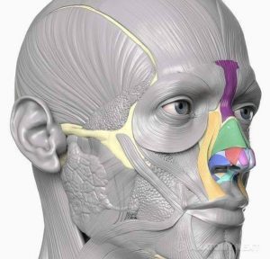
The opposite function (eye opening) is due to the synergistic action of the levator palpebrae superioris muscle, which "lifts" the eyelid like a blind as the orbicularis oculi relaxes, allowing the lower eyelid to drop almost by gravity.
It is important to remember that the levator palpebrae superioris is not considered a facial muscle, as it originates inside the orbit and ends in the upper eyelid; furthermore, its innervation depends on the III cranial nerve (oculomotor nerve), unlike the facial muscles, whose innervation is the responsibility of the VII cranial nerve (facial nerve).
Muscles of the nose
Of these muscles, only one (procerus muscle) is involved in expression, while the rest have a specific function in the respiratory system.
- Procerus muscle
- Transverse nasalis.
- Myrtiformis.
- Dilator nasi.
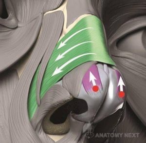
The myrtiformis muscle is responsible for "depressing" the nasal ala and thus closing the entrance of the nostrils, which is particularly useful for limiting the entry of impurities such as dust into the upper respiratory tract.
The transverse and dilator nasi muscles act synergistically to do the opposite: dilate the entrance of the nostril to allow air to enter more easily.
Generally, their action is not visible except in cases of severe respiratory distress, when their effect is so evident that it gives rise to a clinical sign known as nasal flaring, which consists of the elevation of the ala of the nose with each inspiration.
Muscles of the mouth and lips
They are the most numerous and most widely distributed, occupying more than 60% of the total area of the face.
These muscles are responsible for most facial expressions. In addition, some aid in phonation, and a few very particular ones allow mastication: the masseter muscles.
- Buccinator.
- Orbicularis oris.
- Levator labii superioris alaeque nasi.
- Levator labii superioris.
- Caninus muscle.
- Zygomaticus major.
- Zygomaticus minor.
- Risorius.
- Depressor anguli oris.
- Depressor labii inferioris.
- Mentalis.
- Masseter.
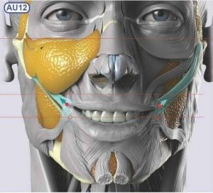
All these muscles, almost entirely innervated by the facial nerve, are responsible for the hundreds of facial expressions of the human face.
For example, a smile is a consequence of the contraction of the risorius and the zygomaticus major and minor muscles; similarly, a slight contraction of this pair of muscles allows the labial commissure to be elevated.
The buccinator retracts the labial commissures; this allows one to whistle, play wind instruments, and dislodge food that accumulates in the vestibular area of the gums.
The orbicularis oris is another specialized muscle that allows the mouth to close, in addition to assisting in the complex movements necessary for sucking.
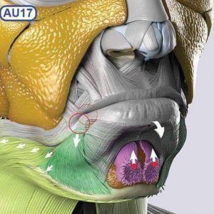
Finally, there is the masseter which, together with the pterygoid muscles (belonging to the pterygoid fossa), forms part of the masticatory muscles.
It is capable of exerting a pressure of 90 kg/cm2, which makes it one of the strongest muscles in the body considering the size-to-force ratio.
It inserts into the zygomatic arch and the ascending ramus of the mandible, allowing for mouth closure and chewing.
To do this, they work in synergy with the rest of the masticatory muscles and in coordination with the neck muscles responsible for mouth opening (digastric, mylohyoid, infrahyoid muscles, among others).
Author Information
- Pessa, J. E., Zadoo, V. P., Adrian, J. E., Yuan, C. H., Aydelotte, J., & Garza, J. R. (1998). Variability of the midfacial muscles: analysis of 50 hemifacial cadaver dissections. Plastic and reconstructive surgery, 102(6), 1888-1893.
- Gasser, R. F. (1967). The development of the facial muscles in man. Developmental Dynamics, 120(2), 357-375.
- Goodmurphy, C. W., & Ovalle, W. K. (1999). Morphological study of two human facial muscles: orbicularis oculi and corrugator supercilii. Clinical Anatomy, 12(1), 1-11.
- Szentágothai, J. (1948). The representation of facial and scalp muscles in the facial nucleus. Journal of Comparative Neurology, 88(2), 207-220.
- Freilinger, G., Gruber, H., Happak, W., & Pechmann, U. (1987). Surgical anatomy of the mimic muscle system and the facial nerve: importance for reconstructive and aesthetic surgery. Plastic and reconstructive surgery, 80(5), 686-690.
- Rubin, L. R., Mishriki, Y., & Lee, G. (1989). Anatomy of the nasolabial fold: the keystone of the smiling mechanism. Plastic and reconstructive surgery, 83(1), 1-10.
- Schwarting, S., Schröder, M., Stennert, E., & Goebel, H. H. (1984). Morphology of denervated human facial muscles. Orl, 46(5), 248-256.